In 2012 the Materials and Condensed Matter Physics Group opened the MagTEM facility at the University of Glasgow. MagTEM is a JEOL ARM (Atomic Resolution Microscope) 200cF which is a scanning transmission electron microscope (STEM) with aberration correction provided by CEOS. Aberration correction allows world leading performance of this instrument, and the capability of the instrument is summarised below:
- standard mode STEM imaging achieves atomic imaging with a spatial resolution of 0.078 nm
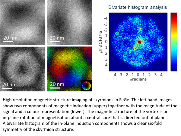
- world’s first magnetic STEM imaging in Lorentz mode with sub nanometre resolution demonstrated [1]
- spectroscopic chemical mapping (electron energy loss spectroscopy (EELS)) can be performed with a Gatan Quantum 965 GIF
- energy dispersive spectroscopy (EDS) with Bruker XFlash 60 X-ray detection system
- in-situ capabilities are provided via specialised and customised sample rods allowing cooling/heating, electrical current application and tomographic reconstruction.
MagTEM has enabled the group to diversify its research base and currently provides support for research projects funded at a level of >£4M, including the recent award of a noble gas FIB (focused ion beam) instrument for improved TEM sample preparation and novel nanopatterning.
In summary MagTEM has become an essential tool across nano-materials science and has enabled collaborations including high resolution studies of a variety of magnetic systems [2,3,4], functional oxides [5], organic electronics [6], and the origins of nanoparticles implicated in the onset of Alzheimer's disease [7].
Further information about the activities of the MCMP group and enquiries regarding the use of the Kelvin Nanocharacterisation Facilities can be found at: http://www.gla.ac.uk/schools/physics/research/groups/mcmp/


[1] Ultramicroscopy 152 (2015) 57
Aberration corrected Lorentz scanning transmission electron microscopy,
S McVitie, D McGrouther, S McFadzean, DA MacLaren, KJ O’Shea and MJ Benitez
[2] Nature Comm. 6 (2015), 8957
Magnetic microscopy and topological stability of homochiral Néel domain walls in a Pt/Co/AlOx trilayer,
MJ Benitez, A Hrabec, AP Mihai, TA Moore, G Burnell, D McGrouther, CH Marrows & S McVitie
[3] New J. Phys. 18 (2016), 095004
Internal structure of hexagonal skyrmion lattices in cubic helimagnets
D McGrouther, R J Lamb, M Krajnak, S McFadzean, S McVitie, R L Stamps, A O Leonov, A N Bogdanov and Y Togawa
[4] Nature 524 (2015) 69
Beating the Stoner criterion using molecular interfaces,
F Al Ma’Mari, T Moorsom, G Teobaldi, W Deacon, T Prokscha, H Luetkens, S Lee, GE Sterbinsky, DA Arena, DA MacLaren, M Flokstra, M Ali, MC Wheeler, G Burnell, BJ Hickey and O Cespedes.
[5] NanoLetters 16 (2016) 5291
Dynamical torque in CoxFe3-xO4 nano-cube thin films characterized by femtosecond magneto-optics: a π-shift control of the magnetization precession,
M Vomir, R Turnbull, I Birced, P Parreira, DA MacLaren, SL Lee, P André and J-Y Bigot.
[6] J. Mater. Chem. C 2 (2014) 245
Structure–function relations in diF-TES-ADT blend organic field effect transistors studied by scanning probe microscopy,
AB Naden, J Loos and DA MacLaren.
[7] PNAS 113 (2016) 10797
Magnetite pollution nanoparticles in the human brain,
BA Maher, IAM Ahmed, V Karloukovski, DA MacLaren, PG Foulds, D Allsop, DMA Mann, R Torres-Jardón and L Calderon-Garciduenas